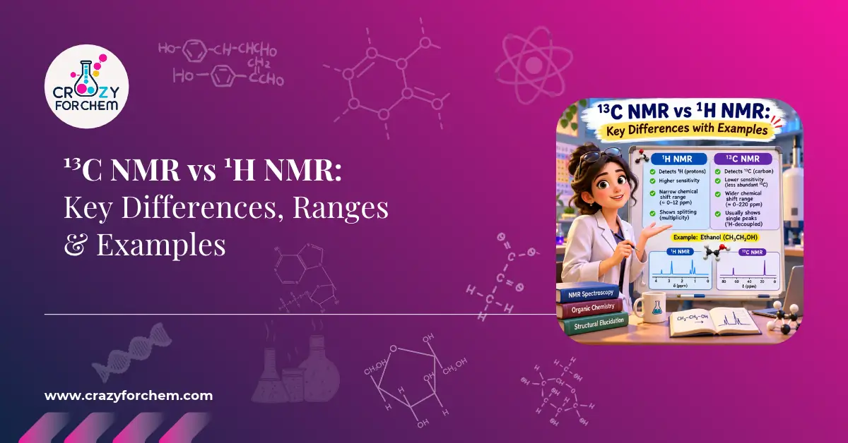
¹³C NMR vs ¹H NMR: Key Differences, Ranges & Examples
Both techniques probe I = ½ nuclei, but ¹H NMR maps the peripheral hydrogen environments and coupling, while ¹³C NMR maps the carbon skeleton directly. ¹³C is about 6,000× less sensitive (1.1% abundance, γ ≈ ¼ of ¹H), spans a far wider 0–220 ppm window, gives sharp singlets under broadband decoupling, and is non-quantitative because of T₁ and NOE effects.
🧪 Click the card to explore
Both ¹H and ¹³C are I = ½ nuclei. ¹H NMR shows how many hydrogens there are, their environments and their neighbours (via J-coupling). ¹³C NMR shows the underlying carbon skeleton — including quaternary carbons that ¹H NMR cannot see. Used together, they give unambiguous structure assignments.
¹H is ~99.98% abundant; ¹³C is only 1.1%. The common isotope ¹²C has I = 0 and is NMR-inactive. The gyromagnetic ratio of ¹³C (67.2 × 10⁶ rad s⁻¹ T⁻¹) is roughly one-fourth of ¹H (267.5 × 10⁶).
Sensitivity ∝ abundance × γ³
Per nucleus: (¼)³ = 1/64 of ¹H
Overall ≈ 1/6000 of ¹H per unit sample
¹H: 0–14 ppm (vs TMS) — a compressed window, so signals often overlap into crowded multiplets.
¹³C: 0–220 ppm (up to ~240 ppm for some carbonyl derivatives) — roughly 20× wider, so almost every non-equivalent carbon gets its own line. Hybridisation, electronegativity and paramagnetic shielding have a much larger effect on carbon shifts.
¹H signals split into multiplets by the n+1 rule (JHH). In ¹³C NMR, ¹JCH (125–250 Hz) would create messy multiplets, so routine spectra use broadband ¹H decoupling — every non-equivalent carbon becomes one sharp singlet.
¹³C–¹³C coupling is unobservable at natural abundance: 1.1% × 1.1% ≈ 0.012% chance of two ¹³C bonded together.
¹H: peak area ∝ number of H — reliable proton counting.
¹³C: two effects distort the areas.
1. Long T₁: carbons relax in seconds to minutes; quaternary carbons relax slowest, so they partially saturate and give shorter peaks.
2. NOE: broadband decoupling transfers polarisation from H to C, boosting intensity by up to ~200% (1 + NOEmax), depending on attached H.
Typical intensity order: CH₃ > CH₂ > CH ≫ C(quaternary)
¹H NMR — 5 signals: CH₃ (C1) 3H doublet δ 1.1; CH₃ (C4) 3H triplet δ 0.9; CH₂ 2H multiplet δ 1.4; CH–OH 1H multiplet δ 3.7; OH broad singlet δ 2.0–4.0.
¹³C NMR — 4 singlets: C4 δ 10.1; C1 δ 22.6; C3 δ 32.2; C2 (C–O) δ 69.3, deshielded by oxygen.
¹H: ring protons sit in a tight δ 6.5–8.5 window, so overlapping multiplets make substitution patterns hard to read.
¹³C: aromatic carbons spread over δ 110–160. Quaternary carbons, such as the O-bearing C1 of phenol (δ ~155), appear as noticeably shorter singlets — long T₁ and weak NOE.
¹H NMR: rapid routine analysis, reaction monitoring, purity checks, neighbour connectivity via J-coupling.
¹³C NMR: carbon backbone, quaternary carbons, polymers and complex natural-product frameworks.
DEPT-90 shows only CH. DEPT-135 shows CH₃ and CH pointing up, CH₂ pointing down, and quaternary carbons absent.
HSQC links each H to its directly attached C (¹JCH). HMBC links H to carbons 2–3 bonds away (²J, ³JCH), tracing the backbone and assigning quaternary carbons.
| Feature | ¹H NMR | ¹³C NMR |
|---|---|---|
| Natural abundance | ~99.98% | ~1.1% |
| Gyromagnetic ratio γ | 267.5 × 10⁶ | 67.2 × 10⁶ |
| Relative sensitivity | High (1.00) | Very low (~1/6000) |
| Shift window | 0–14 ppm | 0–220 ppm |
| Primary data | H count, environments, coupling | Carbon skeleton, hybridisation |
| Multiplicity | Multiplets (n+1 rule) | Singlets (decoupled) |
| Integration | Quantitative | Non-quantitative (T₁, NOE) |
| Acquisition time | Seconds to minutes | Minutes to hours |
| Experiment | What it tells you |
|---|---|
| DEPT-90 | Methine (CH) carbons only |
| DEPT-135 | CH₃ / CH up, CH₂ down, C absent |
| HSQC | H to directly attached C (¹JCH) |
| HMBC | H to C two or three bonds away |
In Modern Instrumental Methods in Chemistry, nuclear magnetic resonance stands as the premier analytical tool for molecular structure elucidation.
While ¹H NMR spectroscopy maps the peripheral hydrogen environments and spin-spin connectivity of a molecule, carbon-13 NMR directly uncovers its underlying carbon skeleton.
Understanding the fundamental differences between 13C NMR and 1H NMR enables chemists to combine both techniques for unambiguous structural assignments.
Table of Contents
Fundamental Physical Differences
Isotopic Abundance and Sensitivity
Nuclear magnetic resonance spectroscopy relies on the magnetic properties of atomic nuclei with non-zero spin quantum numbers (I = 1/2). In proton NMR (1H NMR), the target nucleus is protium (1H NMR), which has an overwhelming natural isotopic abundance of approximately 99.98%.
Conversely, in carbon NMR (13C NMR), the magnetically active isotope is Carbon-13 (13C NMR), which has a natural abundance of only 1.1%. The predominant carbon isotope in nature, Carbon-12 (12C), contains an even number of protons and neutrons (I = 0) and is completely NMR-inactive.
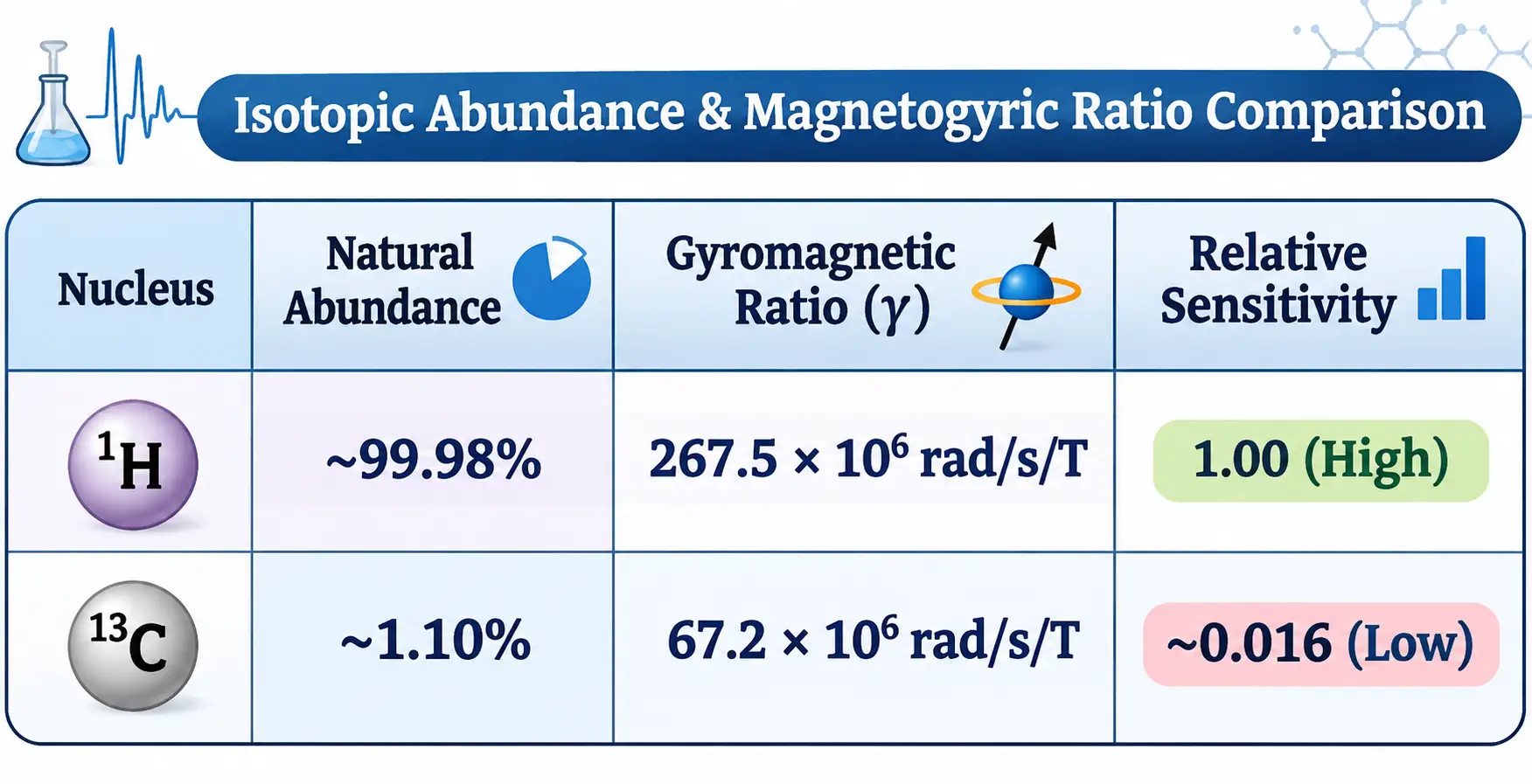
In addition to isotopic scarcity, the gyromagnetic ratio NMR parameter for ¹³C NMR (γ = 67.2 × 10⁶ rad s⁻¹ T⁻¹) is roughly one-fourth that of ¹H (γ = 267.5 × 10⁶ rad s⁻¹ T⁻¹).
Because resonance sensitivity depends on both natural abundance and the cube of the gyromagnetic ratio, an individual ¹³C nucleus yields a resonance signal approximately 1/64th as intense as a proton, making overall ¹³C detection roughly 6,000 times less sensitive than 1H analysis per unit sample.
Understanding this fundamental difference between ¹³C and ¹H NMR explains why Carbon-13 analysis requires Fourier Transform NMR (FT-NMR) systems.
Modern FT-NMR spectrometers apply pulsed radiofrequency excitation to accumulate and average hundreds to thousands of Free Induction Decay (FID) scans, effectively suppressing background noise to yield clean spectral peaks.
Evaluating ¹³C NMR vs ¹H NMR highlights how advanced instrumentation overcomes intrinsic quantum limitations to deliver clear skeletal data.
Chemical Shift Dispersion (0–14 ppm vs 0–220 ppm)
The chemical shift range NMR window for ¹H NMR is comparatively narrow, typically spanning 0 to 14 ppm relative to the internal standard tetramethylsilane (TMS).
Because organic compounds often contain multiple hydrogen atoms in similar electronic environments within this compressed scale, proton signals frequently overlap, giving rise to complex, crowded multiplets.
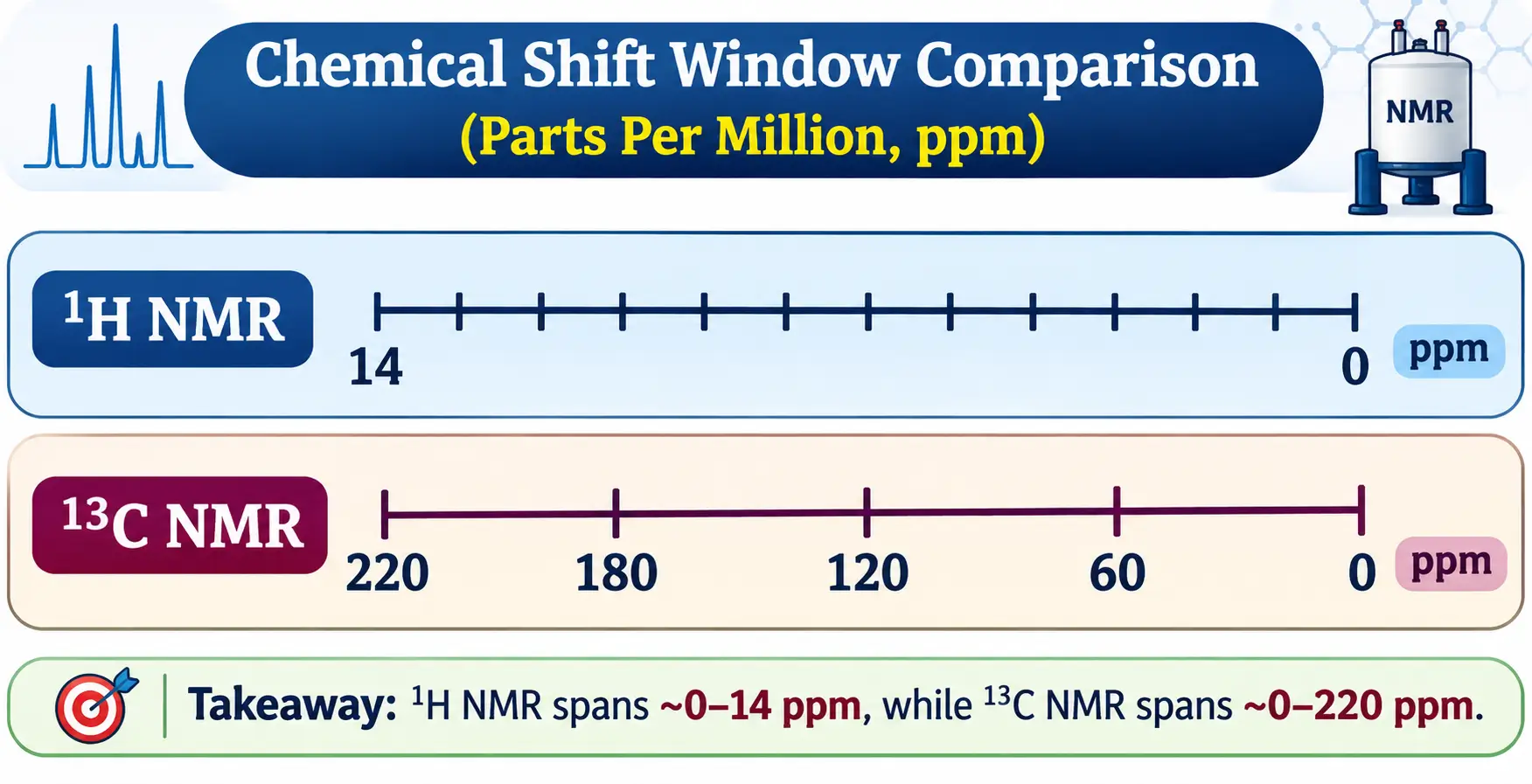
Conversely, the ¹³C NMR chemical shift range is vastly wider, extending from 0 to 220 ppm (and up to 240 ppm for specialised carbonyl derivatives). A useful rule of thumb indicates that ¹³C chemical shifts are roughly 20 times larger than the corresponding 1H shifts for analogous functional groups.
This expanded dispersion ensures that almost every chemically non-equivalent carbon atom in a molecule resonates at a distinct frequency, dramatically reducing peak overlap.
Conducting a direct Carbon-13 vs Proton NMR evaluation shows that while proton spectra reveal peripheral functional environments, carbon spectra cleanly resolve the central molecular architecture.
Spectral Features and Appearance
Multiplicity and Coupling (Spin-Spin Coupling vs Decoupling)
In 1H NMR spectra, scalar J-coupling between adjacent non-equivalent protons causes resonance peaks to split into multiplets (singlets, doublets, triplets, quartets) governed by the (n+1) rule. This splitting provides valuable information regarding neighbour connectivity.
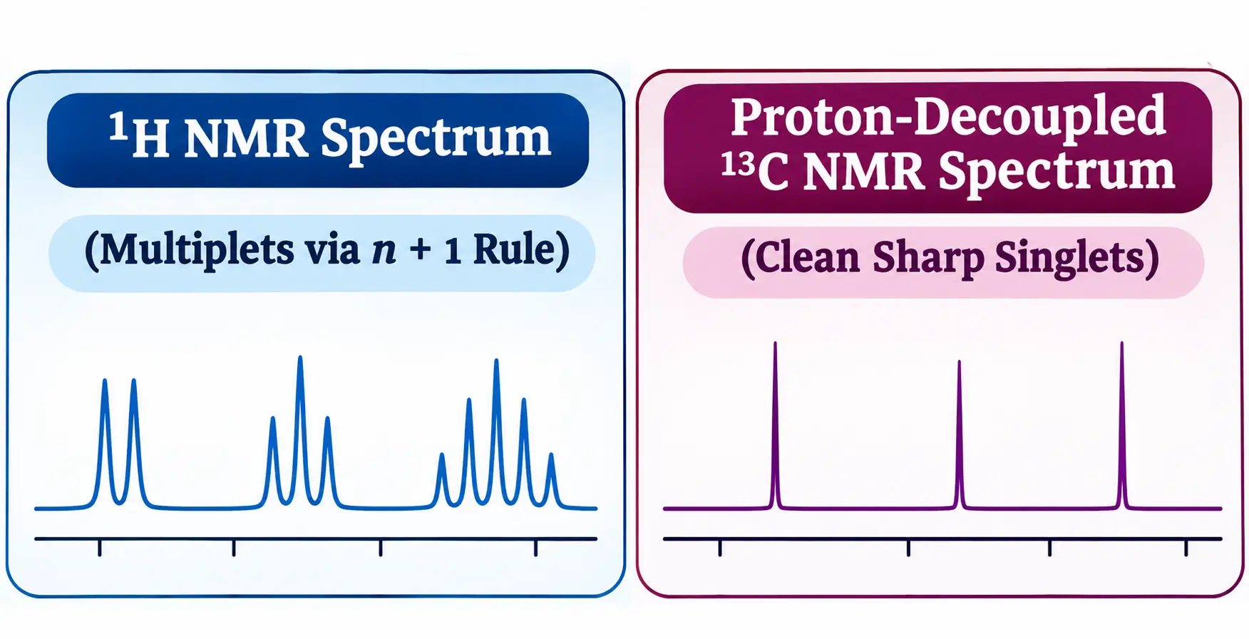
In routine ¹³C NMR, heteronuclear coupling between ¹³C and attached 1H nuclei (¹JCH = 125–250 Hz) would split carbon signals into complex multiplets, severely reducing peak intensity and complicating interpretation.
To prevent this, a standard proton-decoupled ¹³C NMR experiment employs broadband decoupling in ¹³C NMR. By continuously irradiating the sample with a broad radiofrequency range at the proton resonance frequency, all hydrogens are rapidly saturated, completely removing (¹³C-1H) spin-spin interactions. Consequently, every chemically non-equivalent carbon atom appears as a single, sharp line.
Furthermore, homonuclear ¹³C-¹³C coupling is unobservable in natural abundance samples. Because the probability of finding two ¹³C atoms directly bonded to one another is extremely low (1.1% × 1.1% ≈ 0.012%), homonuclear splitting is statistically absent from standard spectra.
Signal Area and Integration Accuracy
A key distinction when analysing ¹³C NMR and ¹H NMR lies in peak integration:
- ¹H NMR Integration: The area under each proton signal is directly proportional to the relative number of hydrogen atoms contributing to that resonance, allowing precise quantitative proton counting.
- ¹³C NMR Integration: Peak areas in standard FT-NMR carbon spectra cannot be directly integrated to determine carbon ratios. This non-quantitative behaviour stems from two physical mechanisms:
- Variable Relaxation Times (T1): Carbon nuclei exhibit long spin-lattice relaxation times (T1) ranging from seconds to minutes. Quaternary carbons (lacking directly attached protons) relax much slower than protonated carbons, leading to partial saturation and diminished peak heights under rapid pulse repetitions.
- Nuclear Overhauser Effect in 13C NMR (NOE): Broadband proton decoupling transfers spin polarisation through dipole-dipole interactions from irradiated protons to adjacent carbon nuclei. This cross-polarisation enhances carbon signal intensities by up to 200% (1 + NOEmax). Because NOE enhancement depends on the number of directly attached hydrogens,
(CH₃ > CH₂ > CH >> Cquat)peak areas are non-uniform across different carbon types.
Comparative Summary Table
The following table provides a comprehensive 13C NMR vs 1H NMR comparison with examples highlighting operational parameters:
| Analytical Feature | 1H NMR Spectroscopy | 13C NMR Spectroscopy |
|---|---|---|
| Observed Nucleus | Protium (1H), (I = 1/2) | Carbon-13 (13C), (I = 1/2) |
| Natural Isotopic Abundance | ~99.98% | ~1.108% |
| Gyromagnetic Ratio (γ) | 267.5 × 10⁶ rad s⁻¹ T⁻¹ | 67.2 × 10⁶ rad s⁻¹ T⁻¹ (~1/4th of ¹H) |
| Relative Detection Sensitivity | High (1.00) | Very Low (~0.016 / ~1/6000th of ¹H) |
| Chemical Shift Window | 0 to 14 ppm | 0 to 220 ppm |
| Primary Structural Data | Proton count, environments, neighbour coupling | Carbon-skeletal backbone, hybridisation |
| Signal Multiplicity | Split into multiplets via the (n+1) rule (JHH) | Single sharp lines (proton decoupled) |
| Homonuclear Coupling | Observed between non-equivalent protons | Unobservable (¹³C–¹³C probability ~0.012%) |
| Peak Integration | Strictly quantitative (proportional to proton count) | Non-quantitative (distorted by (T1) relaxation & NOE) |
| Acquisition Time | Fast (seconds to minutes) | Slower (minutes to hours; requires FT signal averaging) |
Examining Carbon-13 vs proton NMR parameters side-by-side demonstrates how their complementary nature provides a complete picture of molecular architecture.
Practical Examples in Structure Elucidation
Example 1: Aliphatic Compound Analysis (2-Butanol)
Analysing 2-butanol (CH₃-CH(OH)-CH₂-CH₃) highlights key differences between ¹³C NMR and ¹H NMR:
                       Structure of 2-Butanol
                                     OH
                                     │
                     CH₃ ─── CH ─── CH₂ ─── CH₃
                     (C1)         (C2)         (C3)         (C4)
- ¹H NMR Spectrum: Displays 5 distinct proton signals with complex splitting and integration ratios:
- C1 methyl protons (-CH₃): 3H doublet at δ 1.1 ppm (J coupled to C2-H).
- C4 methyl protons (-CH₃): 3H triplet at δ 0.9 ppm (J coupled to C3-H₂).
- C3 methylene protons (-CH₂-): 2H multiplet at δ 1.4 ppm.
- C2 methine proton (-CH-OH): 1H multiplet downfield at δ 3.7 ppm.
- Hydroxyl proton (-OH): 1H broad exchangeable singlet at δ 2.0–4.0 ppm.
- ¹³C NMR Spectrum: Displays 4 distinct singlet lines corresponding to the 4 non-equivalent carbons without splitting:
- C1 methyl carbon (C1): Singlet at δ 22.6 ppm.
- C4 methyl carbon (C4): Singlet at δ 10.1 ppm.
- C3 methylene carbon (C3): Singlet at δ 32.2 ppm.
C2 oxygen-bearing carbon (C-O): Deshielded singlet downfield at δ 69.3 ppm.
Example 2: Substituted Aromatic Analysis (Phenol & Toluene)
Evaluating aromatic derivatives illustrates how comparing 13C NMR vs 1H NMR resolves structural overlap:
- 1H NMR Spectrum: Aromatic ring protons resonate in a tight window between δ 6.5 and 8.5 ppm. Overlapping multiplets frequently make it difficult to determine exact substitution patterns without 2D techniques.
- 13C NMR Spectrum: Aromatic carbons span a wide range from δ 110 to 160 ppm. Quaternary aromatic carbons (such as the oxygen-bearing C1 of phenol
δ 155 ppm) appear as noticeably shorter singlet lines due to longer T1 relaxation times and reduced NOE enhancement. In contrast, 1H NMR cannot directly detect quaternary carbons.
Advanced Techniques and Complementary Usage
When to Use ¹H NMR vs ¹³C NMR
- ¹H NMR: Ideal for rapid routine analysis, real-time reaction monitoring, checking sample purity, and establishing neighbour connectivity via J-coupling.
- ¹³C NMR: Essential for establishing the underlying carbon backbone, identifying quaternary carbon centres, characterising polymers, and resolving complex natural product frameworks.
2D NMR as the Ultimate Bridge (HSQC and HMBC)
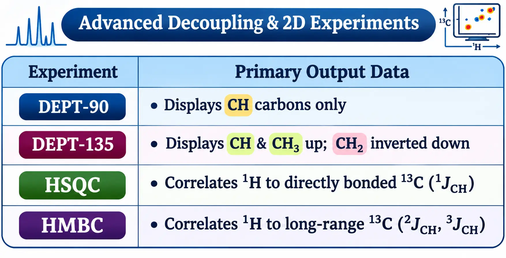
Evaluating DEPT NMR vs ¹³C NMR (Distortionless Enhancement by Polarisation Transfer) provides a fast 1D method to classify carbon sub-types: DEPT-90 isolates methine (CH) carbons, while DEPT-135 displays methyl (CH₃) and methine (CH) carbons pointing up, methylene (CH₂) carbons inverted downwards, and quaternary carbons nulled.
To bridge one-dimensional spectra, researchers utilize 2D heteronuclear correlation experiments:
- HSQC (Heteronuclear Single Quantum Coherence): Correlates each proton signal to its directly attached carbon atom (¹JCH), utilising sensitive proton detection to map single bonds.
- HMBC (Heteronuclear Multiple Bond Correlation): Connects protons to carbons separated by 2 or 3 bonds (²JCH, ³JCH), enabling researchers to trace the entire carbon backbone and assign quaternary carbons with complete confidence.
By deploying ¹H NMR, ¹³C NMR, DEPT, and 2D correlation experiments in tandem, chemists establish robust, unambiguous molecular structure assignments.
Frequently Asked Questions
What is the difference between ¹³C NMR and ¹H NMR?
While both techniques probe nuclei with a spin quantum number of I = 1/2, ¹H NMR analyses the peripheral hydrogen atoms of a molecule, whereas ¹³C NMR directly maps the underlying carbon skeleton. Key operational differences include natural isotopic abundance (99.98% for ¹H vs 1.1% for ¹³C), chemical shift range (0–14 ppm vs 0–220 ppm), peak integration accuracy, and spin-spin splitting behaviour.
Why is ¹³C NMR less sensitive than ¹H NMR?
¹³C NMR is roughly 6,000 times less sensitive per unit sample than ¹H NMR due to two physical factors: its low natural isotopic abundance (1.1% compared to 99.98% for ¹H, as ¹²C is NMR-inactive) and its smaller gyromagnetic ratio (γ), which is approximately one-fourth that of a proton. Consequently, ¹³C NMR requires Fourier Transform (FT-NMR) signal averaging over hundreds of scans to achieve a clear signal-to-noise ratio.
What are the two main types of NMR spectroscopy used in organic chemistry?
In routine organic structure elucidation, the two primary types of 1D nuclear magnetic resonance spectroscopy are Proton NMR (¹H NMR) and Carbon-13 NMR (¹³C NMR). Used in tandem, ¹H NMR establishes proton counts and neighbouring connectivity via J-coupling, while ¹³C NMR identifies carbon hybridisation, functional groups, and quaternary carbon centres.
Why are there no splitting patterns (multiplets) in a standard ¹³C NMR spectrum?
Routine ¹³C NMR spectra are acquired using broadband proton decoupling, which continuously irradiates all protons to eliminate heteronuclear ¹³C–¹H splitting and collapse signals into sharp singlets. Furthermore, homonuclear ¹³C–¹³C coupling is statistically absent because the probability of two low-abundance ¹³C isotopes sitting adjacent to each other is only around 0.012%.
Why can peak areas not be integrated in ¹³C NMR like they are in ¹H NMR?
Unlike ¹H NMR—where peak integrals are strictly proportional to proton count—standard ¹³C NMR peak intensities are non-quantitative due to varying spin-lattice relaxation times (T₁) and the Nuclear Overhauser Effect (NOE). Quaternary carbons relax slowly and receive minimal NOE enhancement, resulting in noticeably shorter peaks than proton-bearing CH, CH₂, or CH₃ carbons.
Why is the chemical shift range of ¹³C NMR (0–220 ppm) much wider than ¹H NMR (0–14 ppm)?
Carbon nuclei possess p-orbitals and valence electrons located much closer to the nucleus than the 1s spherical electron cloud of hydrogen. Changes in hybridisation, electronegativity, and paramagnetic shielding therefore exert a roughly 20-fold larger effect on carbon chemical shifts, spreading resonances across a wide 0–220 ppm window that minimises spectral overlap.

Founder · CrazyForChem
Chemistry Educator | Organic Chemistry Expert | M.Sc. Organic Chemistry
Helping Students & Teachers Master Chemistry. I’m a passionate chemistry educator who loves making chemistry simple, relatable, and fun. With a background in Organic Chemistry, I help students not only understand concepts but also explore the many career paths chemistry can lead to.